Introduction
The comprehensive characterization of a wide range of analytes and samples using Asymmetrical Flow Field-Flow Fractionation (AF4) coupled with various detection techniques has been demonstrated in numerous studies. In the present work, we describe the in-line coupling of AF4 with Small-Angle X-ray Scattering (SAXS) to enable detailed characterization of complex sample compositions. SAXS is a versatile technique for the characterization of pharmaceutically relevant macromolecules, providing information on their absolute concentration, size, shape, and internal structure [1,2].
A prerequisite for reliable SAXS analysis is the availability of highly pure and monodisperse analytes. This limitation can be addressed by coupling SAXS with AF4, which separates sample constituents according to their hydrodynamic size prior to SAXS analysis, thereby enabling size-resolved structural characterization. Here, we present the in-line coupling of multi-detector AF4 with SAXS detection, allowing quantitative, multi-attribute analysis of mRNA nanoparticles within a single measurement.
Experimental
A mRNA lipoplex sample (LPX) provided by BioNTech was chosen as a test sample. It contained negatively charged, polydisperse LPX nanoparticles and free unbound mRNA and was obtained by self-assembly between cationic liposomes and mRNA (Figure 1). The carrier liquid for AF4 separation was a HEPES buffer. For fractionation, a semi-preparative AF4 frit-inlet channel with a channel height of 350 μm and equipped with a 10 kDa polyethersulfone membrane was used. The frit-inlet channel was also equipped with the slot outlet function Io increase the analyte concentration prior to detection. For connecting the AF4-UV-MALS system to SAXS a 1 mm quartz flow capillary in a ´flow-through mode´ was used. SAXS data was collected at the P12 bioSAXS beamline of the European Molecular Biology Laboratory at the PETRA III synchrotron radiation source, DESY Hamburg (Figure 2). The samples were injected as received.

Figure 1: Schematic of the LPX structure [1].
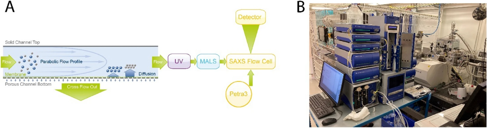
Figure 2: Schematic overview of the multi-detector AF4 setup (A) and picture of the instrumental setup at the EMBL P12 bioSAXS beamline used in this study (B) [1].
Results
Figure 3 shows the AF4-UV-MALS-SAXS fractogram of the investigated LPX sample. Efficient AF4 separation was achieved, as evidenced by two distinct fractions eluting at approximately 18 min and 40 min. Analysis of the MALS data enabled determination of the radius of gyration (Rg) and molar mass (MM). The first fraction exhibited a constant Rg of 25 nm and a MM of 475 kDa, consistent with the theoretical value for pure mRNA. The second fraction, assigned to LPX particles, showed a broad Rg distribution ranging from 80 to 470 nm, demonstrating the pronounced polydispersity of the sample.

Figure 3: AF4 fractogram of the LPX sample with UV (purple), 90° MALS (blue), SAXS (yellow) signal & Rg (black) derived from MALS data vs elution time [1]
The successful fractionation of free mRNA from LPX is also illustrated in Figure 4. Compared with the unfractionated (batch) sample, the fractionated LPX sample showed a lower scattering slope toward q=0, confirming the effective separation from free mRNA. The Bragg peak observed in the SAXS scattering curve is characteristic of the lamellar structure of LPX particles, where the mRNA is arranged between repeating lipid bilayers. In addition, the scattering intensity and slope below the Bragg peak indicate compact particles with smooth surfaces. These results highlight the importance of fractionation for obtaining meaningful SAXS data from polydisperse LPX samples. Figure 5 shows the number of mRNA copies in each LPX size fraction, revealing a clear increase in mRNA payload with increasing particle size. The SAXS data also provides additional structural information for each size fraction, including the absolute particle concentration, the total number of particles, the spacing between the lipid layers (d-spacing), and the correlation length. This size-resolved analysis provides a comprehensive picture of the structural properties of LPX particles across the entire size distribution.

Figure 4: SAXS patterns of LPX: batch measurement (grey) and after separation with AF4 (yellow) [1].

Figure 5: Total number of mRNA copies derived from SAXS data in certain particles size fractions (Rg) from MALS (green bars) and mRNA copies per particle of size segment (red dots) [1]
Conclusion
The hyphenation of multi-detector AF4 with SAXS enabled the size-resolved assessment of multiple critical quality attributes of a mRNA lipoplex sample in a single measurement including quantitative information on particle size distribution profiles, drug loading, size-dependent internal structures and free drug. This approach is particularly applicable to complex nanoparticulate structures [3,4] and has the potential to become a standard tool for samples in the size range of 100 nm and below. For more information on the detailed measurement protocols, data evaluation and results we recommend further reading in Graewert et al., 2023, Scientific Reports [1]. This setup is now permanently integrated at the EMBL P12 bioSAXS beamline at the PETRA III synchrotron at DESY, Hamburg, and requests for measurement time are always welcome.
References
[1] M. A. Graewert, C. Wilhelmy, T. Bacic, J. Schumacher, C. Blanchet, F. Meier, R. Drexel, R. Welz, B. Kolb, K. Bartels, T. Nawroth, T. Klein, D. Svergun, P. Langguth, H. Haas, Scientific Reports, 2023, 13, 15764.
[2] S. Da Vela, K. Bartels, D. Franke, D. Soloviov, T. Graewert, D. Molodenskiy, B. Kolb, C. Wilhelmy, R. Drexel, F. Meier, H. Haas, P. Langguth, M. Graewert, Journal of Synchrotron Radiation, 2025, 32, 971-985.
[3] H. Bolinsson, M. C. Pedersen, M. Glantz, F. Herranz-Trillo, J. J. K. Kirkensgaard, L. Nilsson, Food Hydrocolloids, 2025, 32, 111377.
[4] P. Knappe, L. Boehmert, R. Bienert, S. Karmutzki, B. Niemann, A. Lampen, A. F. Thünemann, Journal of Chromatography A, 2011, 1218, 4160-4166